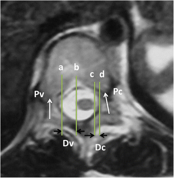
Fig. 2

The measurement of the shift of spinal cord. The transverse plane of apex (T8) in MRI with T2 weighted imaging for a female with idiopathic scoliosis (right thoracic scoliosis, a Cobb’s angle 46°, Lenke 1C). Pv and Pc represent direction of pedicle on the convex and concave side of apex. Line a and b, line c and d were level with Pv and Pc respectively. Line a and b were tangents of medial wall of pedicle and spinal cord on the convex side of the apex. Line c and d were tangents of medial wall of pedicle and spinal cord on the concave side of the apex. The vertical distance between line a and b (Dv) and line c and d (Dc) represented distance between spinal cord and medial wall of pedicle on the convex and concave side respectively